Machine learning detects early signs of kidney damage
Artificial intelligence systems have been taught to detect abnormal cell structures invisible to the human eye, potentially speeding up diagnosis of kidney failure.
Acute kidney injury (AKI) imposes a significant financial burden on healthcare systems, considering the number of hospitalizations and other associated costs.
Luckily, machine learning techniques could change all this by detecting subtle signs of kidney damage, potentially enabling early intervention and treatment before they turn into serious problems.
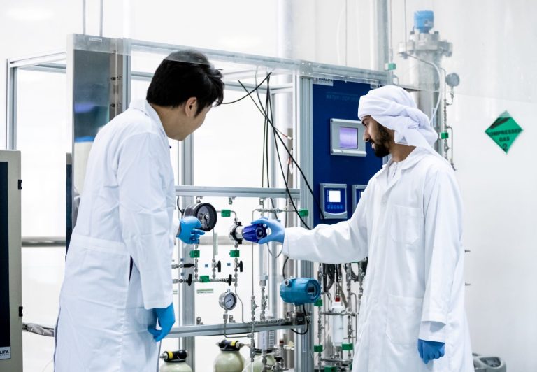
Researchers at Khalifa University working with international colleagues have shown that artificial intelligence (AI) systems can detect abnormal cell structures that are virtually imperceptible to the human eye. Using existing biopsy materials taken from patients the technique could speed up diagnosis of kidney failure in hospitals.
There are differences in the onset of this type of injury that we would not be able to detect, whereas the machine can
Peter Corridon
“There are differences in the onset of this type of injury that we would not be able to detect, whereas the machine can,” says Peter Corridon, who specializes in kidney physiology and who led the project.
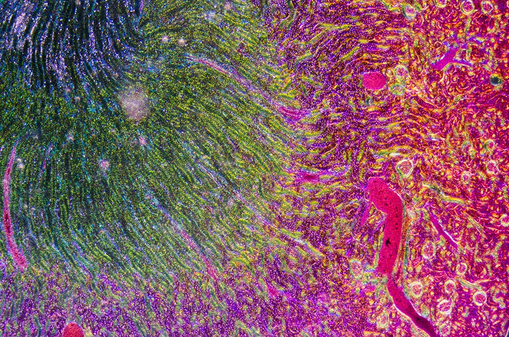
To test the AI’s ability to detect kidney damage, the researchers showed the computer images of cells from rat organs. Some images showed healthy tissue and others showed damage to proximal tubule cells, the most common cell type in the kidney, which deals with metabolic waste products. Damage to these cells is associated with the abrupt drop in renal function seen in AKI and other hard-to-treat and potentially life-threatening consequences.
When provided with information about which tissue images were taken from animals with kidney injury, the AI was able to find signature changes in the shape and structure of the proximal tubule cells associated with the pathology.
The study could pave the way for the development of a “sensitive, accurate, and affordable computer-aided diagnostic system that might be an essential addition to current nephropathology practices,” the researchers say.
One reason AKI is hard to diagnose lies in the limitations of conventional microscope images of kidney tissues taken in biopsies, which are not detailed enough to pick up small changes in cell nucleus structure. To improve the quality of the scans, the researchers used innovative techniques called gray -level co-occurrence matrix analysis and discrete wavelet transform. These methods generate images with increased texture, providing the AI with more features and patterns to associate with the disease state.
The next step for the research team is to test the AI system with more unlabelled images, allowing it to distinguish between healthy and diseased tissue. “Now we are working with a pathologist and doing this in a blind formation,” Corridon says. “So having the pathologist tell us this, and the machine tell us this, and seeing who is correct.”
References
- Pantic, I., Cumic, J., Dugalic, S. et al. Gray level co-occurrence matrix and wavelet analyses reveal discrete changes in proximal tubule cell nuclei after mild acute kidney injury. Scientific Reports 13, 4025 (2023). | Article


